Several years ago, when this disease was at it's worst and affecting all of my trees and shrubs,
this tree appeared to be the least affected.
So, when I first started to scan a branch of this tree, I assumed I wouldn't find many spores.
I was wrong - there was an abundance of spores, leading me to conclude that while it too was heavily
infected, it didn't show its infection as readily through distorted leaf growth as did other
trees and shrubs.
One of the attractions of a Japanese Stewartia is its unusual characteristic of having
naturally exfoliating (peeling) bark.
However, a major characteristic of white canker is split and peeling bark.
Therefore, it's particularly hard to diagnose this disease on a Japanese Stewartia by
its lack of intact bark or distorted leaves. The tree may look healthy, but be badly diseased,
as the following pictures show.
This particular Japanese Stewartia tree is about 10' high.
1
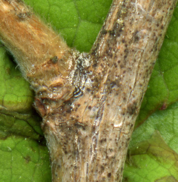
This branch section was about 8" from the branch tip, and about 4' off the ground.
It has a relatively small spore density - all spores are concentrated at the branch junction.
There are also dark brown spots in close proximity to the spores.
2
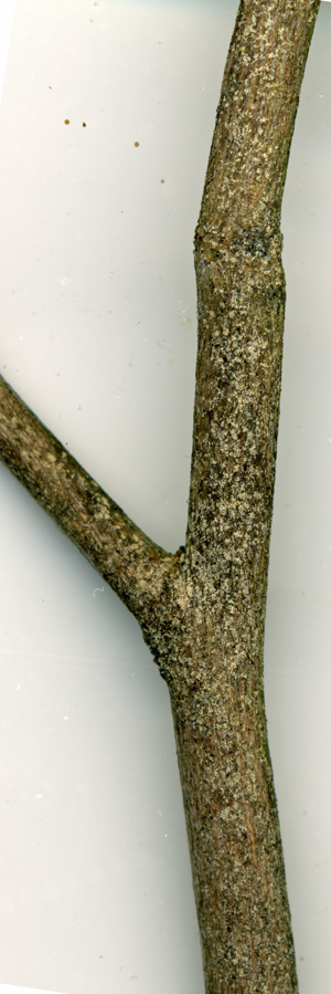
This branch segment, about 2' in from the branch tip, also shows an abundance of spores
at the branch junction.
Nearby junctions also have a high spore density.

3
While not as numerous, the bark between junctions also has a significant spore density, as
this picture shows.
Strangely, when I flipped this branch over to scan the top side, I saw virtually no spores at all!
But I did see a few bark splits and dark spots.
The dark brown spots were mainly at the branch junction, hinting that they are associated with
infected areas.
4
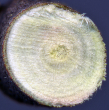
This is a cross-section of the 1/4" branch shown in picture 3.
Visually, it appears healthy. That's because the damage is mainly microscopic, as the
next pictures show.
The above pictures were taken in early May, 2008, using a scanner. The following pictures were taken
two months later, in early August, using a 400 power microscope.
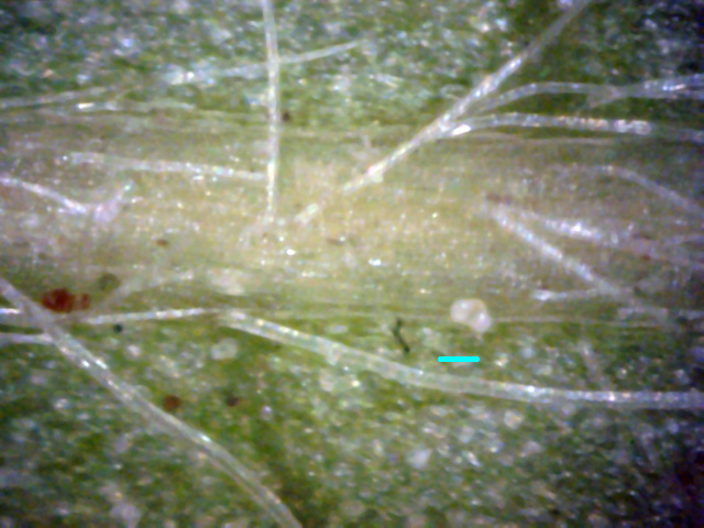
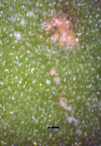
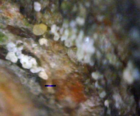
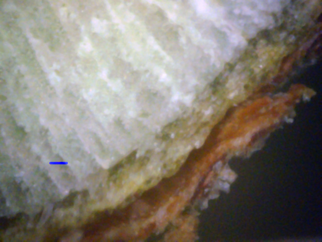
The short blue lines in all the pictures are 50μm scale bars.
This is the approximate diameter of a white canker spore.
5
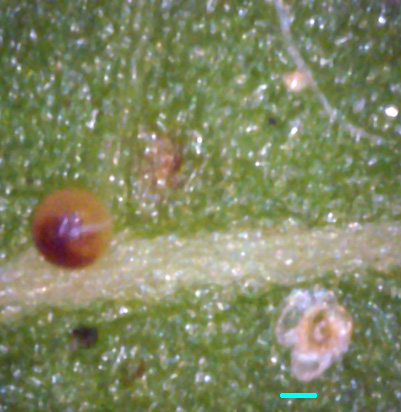
This is an area around the stem on the bottom of the leaf.
The vein hairs are normal.
There is only a single spore showing - its above the 50μ blue leaf scale bar.
The remainder of the leaf bottom is free of any sign of disease.
6
These two leaftop pictures show almost a complete absence of any disease symptoms.
The faint white spots in picture 6 are likely white canker blobs within the leaf interior.
There appears to be a buried hypha and spore in picture 7 (black arrow).
The brown spherical object in picture 7 is unknown.
8
Looking over the bark of a twig microscopically, there was again little evidence of infection.
While this is at odds with pictures 1, 2, and 3, those pictures were taken in late May, about
a week before a major spore release. These microscope pictures were taken in early August, two months
after the spore release.
In spite of this, I did find one pocket of spores (shown here) located at the junction of a twig and
its parent branch. This junction is where white canker spores usually are found.
Note, however, that the spores are now milk white, and have lost their youthful yellow tinge.
9
↓
←
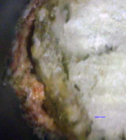
A microscopically examined twig cross-section turned out to be the best indicator of white canker.
Here you can see that the normally green phloem is almost half-consumed with white canker material.
There is also a layer of white canker material within the outer bark (blue arrow), and some on
the surface of the outer bark (red arrow).
10
←
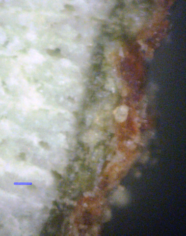
In this cross-section segment, white canker has consumed almost all of the green phloem,
about half the outer bark, and is starting to consume the xylem (sapwood).
Note that the white canker substance is all a uniform gray in color, while the xylem is milk white.
The black arrow shows where the canker found a void and grew into it.
11
→
White canker growth is often diffuse. The exception is where it is forming a spore.
It tends to form spores in an internal void (a pretty useless strategy),
in the outer bark (also useless), or on the surface of the outer bark (far more productive).
Here we see a spore growing within the outer bark (black arrow).
There was so much canker growth in the phloem under it that the outer bark was pushed out.
In summary, a visual check of deformed leaves or separating bark will not be helpful in
diagnosing white canker. Instead, examination with a microscope is needed. The best place to
check for white canker spores is on the underside of branch junctions, a foot or two from
the branch tip. Another good way to diagnose white canker is to examine a twig (e.g., 3/16" diameter)
cross-section and check for patches of a gray-white substance within the phloem and within the bark.
If there is a void between the phloem and the outer bark, or if the white canker has invaded the
xylem, then the infection is particularly severe.
